Figure 1.
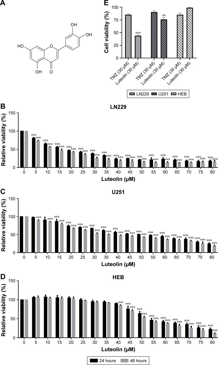
Luteolin inhibited cell proliferation in glioma cells.
Notes: (A) The molecular structural formula of luteolin. (B) LN229 cells were incubated with different concentrations of luteolin for 24 and 48 hours, and then the cell viability was measured by CCK-8 assay. (C) U251 cells were incubated with different concentrations of luteolin for 24 and 48 hours, and then the cell viability was measured by CCK-8 assay. (D) HEB cells were incubated with different concentrations of luteolin for 24 and 48 hours, and then the cell viability was measured by CCK-8 assay. (E) LN229, U251, and HEB cell lines were incubated with luteolin and temozolomide, respectively, for 24 hours. Data are expressed as the mean ± SD of three independent experiments (**P<0.01, ***P<0.001 vs the control group; n=3).
Abbreviations: CCK-8, Cell Counting Kit-8; TMZ, temozolomide.

