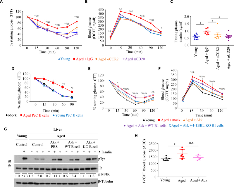
Fig. 7. Gut dysbiosis in aging promotes IR via 4BL cells.
(A) ITTs, (B) OGTTs, and (C) fasting plasma insulin show that IR in aged mice is reversed by the depletion of monocytes and B cells (n = 7 to 8 per group, as in Fig. 2D; “*,” “#,” and “&” show for P < 0.05 for comparisons of the following groups of mice: young versus aged + IgG, aged + IgG versus aged + αCCR2, or aged + αCD20, respectively). (D) Sort-purified or B1a cells from young or aged mice were intravenously transferred into B cell–deficient JHT mice fed with an HFD for 4 months. ITT was performed 1 week after intravenous transfer (n = 10 per group; “#” is for P < 0.05 for comparisons of PeC B cells from young versus aged mice). (E and F) Aged mice were gavaged with A. muciniphila for 20 days, but at day 14, mice were intravenously transferred with sort-purified B1a cells from the PeC of WT or 4–1BBL KO aged mice (Aged + Akk + WT B1 or Aged + Akk + KO B1, respectively) to evaluate IR at days 20 to 22 (n = 8 per group, with the experiment reproduced three times; “*,” “#,” “&,” and “%” are for P < 0.05 for comparisons of the following groups of mice: young versus aged + mock, aged + mock versus aged + Akk, aged + Akk versus aged + Akk + WT B1, or aged + Akk + KO B1, respectively). (G) At day 24, mice in (E) and (F) were injected with insulin to quantify pTyr induction of the liver insulin receptor (as in Fig. 6D). Numbers show the average pTyr increase normalized to β-tubulin. (H) IR was also increased in aged macaques (M. mulatta, n = 6 to 7 per group), which was reversed by Abx treatment (as in Fig. 3, H and I). Data are means ± SEM. *P < 0.05, **P <0.001, and ***P < 0.0001 (Mann-Whitney test and Kruskal-Wallis test with Dunn’s test for multiple corrections).

