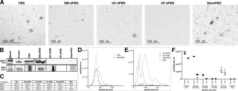
Figure 1.
Characterization of EVs isolated by ultracentrifugation. Transmission electron microscopy analysis revealed the presence of vesicles in FBS, UC-dFBS and SBI-dFBS. StemPRO contained scarce vesicle-like structures and abundant protein aggregates, while no vesicles could be detected in UF-dFBS (A). Western blotting (B) displayed strong expression of CD71 in FBS and SBI-dFBS, no expression in UC-dFBS and UF-dFBS, whereas low expression was detected in StemPRO. Expression of HDL was seen in all samples except UF-dFBS. Nano tracking analysis (C–F) indicated that UF-dFBS had the lowest particle count. Particle distribution (C–F). (C) D10 = 10% of particles are below the size indicated as D10, D50 = 50% of particles are below the size indicated as D50, D90 = 90% of particles are below the size indicated as D90. Median particle size in FBS, dFBS and UC-FBS are in a similar range, while UF-dFBS is considerably different. (D,E) Particle concentration and distribution. FBS and SBI-dFBS showed similar particle concentrations. (F) X-axis; particle size (nm) in two size ranges; 50–200 nm and 200 + nm.

