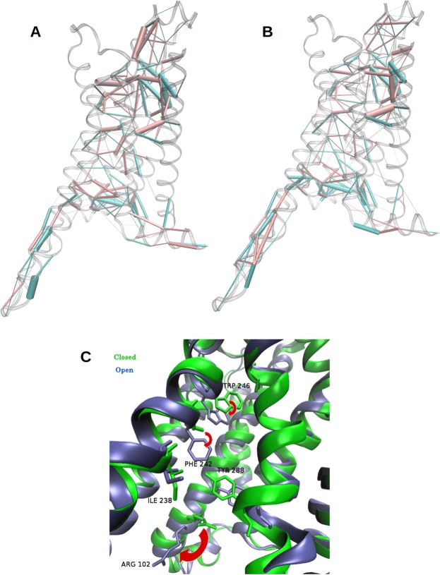
Figure 4.
Rearrangement of contacts in A2aR. (A) Differences between side chain interactions in A2aR and A2aR:Mini-Gs, revealed by analysis of Protein Structure Networks (PSNs). Pink cylinders: predominant contacts in A2aR:Mini-Gs; blue cylinders: predominant contacts in A2aR. The thickness of the cylinders is proportional to the difference in interaction strength. (B) Same analysis for the A2aR:α5 complex. (C) Conformational changes of side chains involved in the redistribution of contacts, that extended from the vicinity of the Mini-Gs binding site to the agonist site. Blue: A2aR; green: A2aR:Mini-Gs. The red arrows indicate the relaxation of W246, F242 and R102 upon removal of Mini-Gs.

