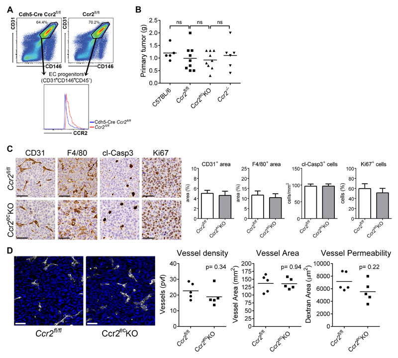
Figure 1. Primary tumor growth was not effected by endothelial deficiency of CCR2.
A) Analysis of endothelial progenitor cells (CD31+CD146+CD45-) in the lungs of naïve Ccr2ecKO mice and Ccr2fl/fl mice. B) Weight of primary subcutaneous LLC1.1 tumors at time of resection (14 days after implantation); ns = not significant. C) Representative images of primary tumors stained with CD31, F4/80, cl-Casp3 and Ki67 Abs together with histological analysis, respectively. Bar = 50 μm. D) Representative images of primary tumors from Ccr2ecKO and Ccr2fl/fl mice, respectively, stained with anti-CD31 (white) to analyze vascular density (nuclear staining with DAPI = blue). The analysis of vessel density and the vessel area revealed no difference between mice of both genotypes. Vessel permeability in tumors was determined using intravenous injection of dextran-FITC 1 h prior to termination. Cryosections of the tumors were analyzed for the dextran-FITC. Bar = 30 μm.

