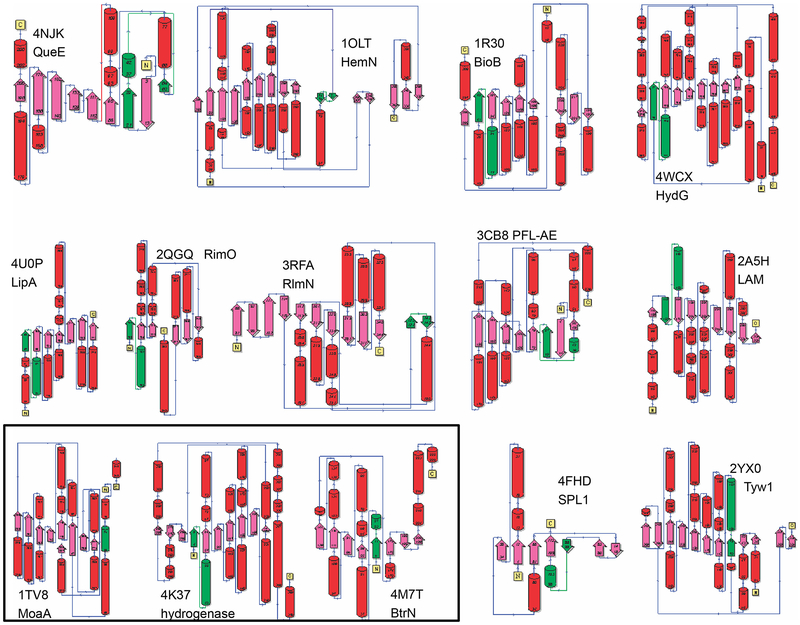
Fig. 6.
Secondary structure topologies of representative radical SAM superfamily domains. Images for several RSS subgroups created using the PDBSum website (de Beer, Berka, Thornton, & Laskowski, 2014). Red: helices, pink: beta strands, green: [Fe4-S4]-AdoMet binding motif. The common abbreviations of the enzyme names and their PDB identifiers are shown on the figure.

