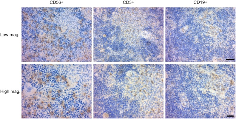
Figure 4. Distribution of splenic T cells, B cells, and NK cells in NSG hL-7xhIL-15 humanized mice.
Thin sections of a hIL-7xhIL-15 NSG recipient spleen stained with anti-hCD56, anti-CD3 and anti-CD19 antibodies. Low and high magnification images are shown. hCD3+ T cell and hCD19+ B cells were found within lymphoid clusters, whereas CD56+ NK cells are located outside the clusters. Scale bars: low magnification, 50 μm; high magnification, 20 μm.

