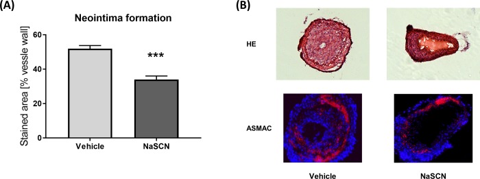
Fig 5. Evaluation of neointima formation after carotid-artery wire injury in wild-type mice upon NaSCN treatment.
(A) Quantitative analysis of neointima formation by use of hematoxylin/eosin staining as a percentage of the vessel wall. (B) Representative histological images of the injured carotid artery 14 days post injury. Upper panel with hematoxylin/eosin staining. Lower panel with anti-α-smooth-muscle Actin staining (red) and DAPI (blue). Data are presented as the mean ± SEM, n = 5, ***p ≤ 0.005 vs. vehicle.

