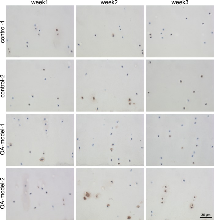
Fig 2. Representative micrographs showing immunohistochemical staining of MMP13 in the middle zone of osteochondral explants over time.
The percentage of positive cells per zone was calculated for each time point, group and horse. Increase of MMP13 positive chondrocytes was detected in OA-model-1 compared to control-1 and in week 3.

