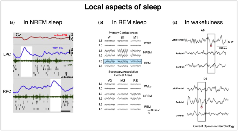
Figure 1.
Local aspects of sleep. (a) Example of local sleep slow waves in human NREM sleep occurring at different times in left and right posterior cingulate cortices, where 100% of units are locked to slow waves. Rows (top to bottom) depict activity in scalp EEG (Cz, red), left posterior cingulate, and right posterior cingulate. Blue, depth EEG; green, MUA; black lines, single-unit spikes. White shadings mark local OFF periods (reproduced, with permission, from Ref. [11]). (b) Laminar recordings revealing slow waves in layer 3 and 4 of mouse primary cortex in REM sleep (reproduced, with permission, from Ref. [14••]). (c) Representative examples of local theta waves (boxed) occurring in left frontal derivations (top panel) after prolonged audio-book (AB) listening and in parietal derivations after playing with a driving simulator game for extended periods of time. Red circles indicate the negative peaks of theta waves detected in each EEG trace (reproduced, with permission, from Ref. [19•]).

