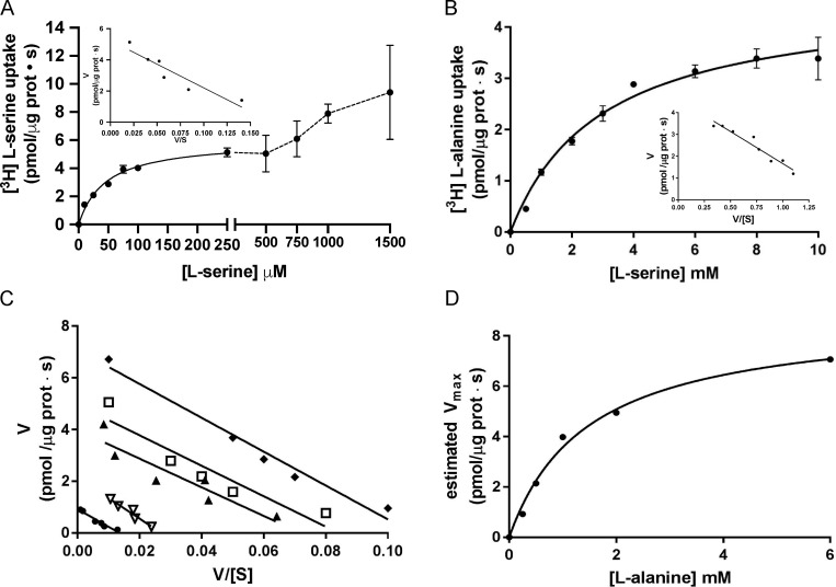
Figure 6.
Kinetic characterization of [3H]L-serine uptake in BasC PLs. (A) Determination of extraliposomal kinetic parameters. Michaelis–Menten plot of the transporter-mediated uptake of [3H]L-serine (1 µCi/µl, 4 s pmol/µg protein · s) in BasC-GFP-PLs containing 4 mM L-alanine, varying extracellular L-serine concentrations (0–1,500 µM). Mediated transport (exchange) was calculated as [3H]L-serine uptake in PLs containing 4 mM L-alanine minus empty PLs. Data correspond to a representative experiment, performed using three replicates. Inset: Eadie–Hofstee plot of the kinetics covering only L-serine concentrations between 0–250 µM (i.e., the high apparent affinity component). Km and Vmax values were 45 ± 5 µM and 6.0 ± 0.2 pmol [3H]L-serine/µg prot · s, respectively. V/[S]: pmol [3H]L-serine/µg prot · s / substrate concentration. (B) Determination of intraliposomal kinetic parameters. Michaelis–Menten plot of the transporter-mediated uptake of [3H]L-alanine (10 µM, 1 µCi/µl, pmol/µg protein · s) in BasC-GFP-PLs containing 0.5–10 mM cold L-serine. Mediated transport was calculated as [3H]L-alanine uptake in L-serine–containing PLs minus empty PLs. Data correspond to a representative experiment, performed using three replicates. Inset: Eadie–Hofstee plot. Km value was 2.5 ± 0.4 mM. In A and B, three independent experiments were performed, giving similar results. (C) Extraliposomal kinetics as in A at different intraliposomal concentrations of L-alanine (0.2, 0.5, 1.0, 2.0, and 6.0 mM). Eadie–Hofstee plots from a representative experiment with the mean values from three replicates. (D) Michaelis–Menten plot of the estimated Vmax values at the different intraliposomal L-alanine concentrations from the kinetic series shown in C.

