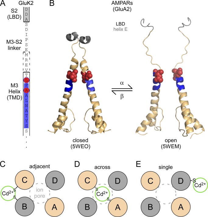
Figure 1.
Opening of the M3 gate. (A) Primary sequence of the upper M3 segment (M3 helix), the linker connecting M3 to the ligand-binding domain (LBD; M3-S2 linker), and the most proximal elements of the LBD (helix E, located in S2). The SYTANLAAF, the most highly conserved motif in iGluRs, is highlighted in blue. Positions A8 and L10 that, when substituted with cysteine, show current potentiation when exposed to Cd2+, are highlighted in red. (B) Structures of the corresponding elements either in the closed state (left, PDB accession no. 5WEO) or in the open state (right, PDB accession no. 5WEM; Twomey et al., 2017). (C–E) Cartoon representations of the possible configurations for coordination of Cd2+ by A8C or L10C. The same subunits (A/C or B/D) are colored the same.

