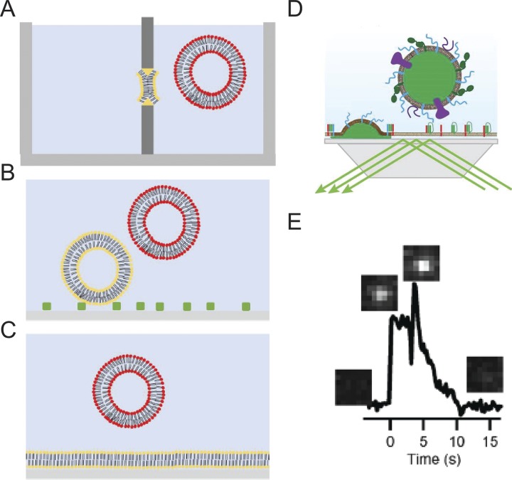
Figure 10.
Assays for reconstitution of membrane fusion. (A) Fusion between a planar membrane (yellow) and liposomes. (B) Vesicle–vesicle fusion where target vesicles (yellow) are tethered to the surface and fusion with secretory mimicking vesicles (red) is imaged. (C) Fusion between target planar supported bilayer (yellow) and secretory mimicking vesicle (red). (D) Cartoon of a “hybrid” granule docking and fusion assay performed using a prism-based TIRF microscope system. (E) A single fusion event of an Neuropeptide (NPY)-Ruby-labeled granule with a planar-supported bilayer containing t-SNAREs. Images corresponding to the appearance of the granule and release of NPY in the evanescent field are placed above an intensity versus time graph. Taken from Kreutzberger et al. (2017), E is reprinted with the permission of Science Advances.

