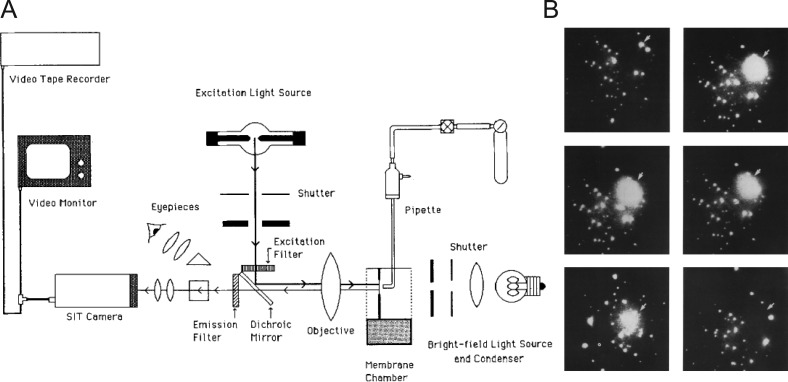
Figure 3.
Fluorescence-based imaging of single-vesicle fusion. (A) Diagram of the fluorescence microscope arrangement and video equipment. The light sources, filters, dichroic mirror, objective, condenser, and eyepieces were components of an upright microscope laid on its backbrace. (B) Individual movie frames from the video record are temporally arranged from left to right and top to bottom. The first panel indicates the time immediately before vesicle rupture and release of calcein (subsequent frames); the intact vesicle is indicated by an arrowhead. The final frame indicates the original location of the vesicle, which is no longer evident. Taken from Niles and Cohen (1987).

