Figure 2.
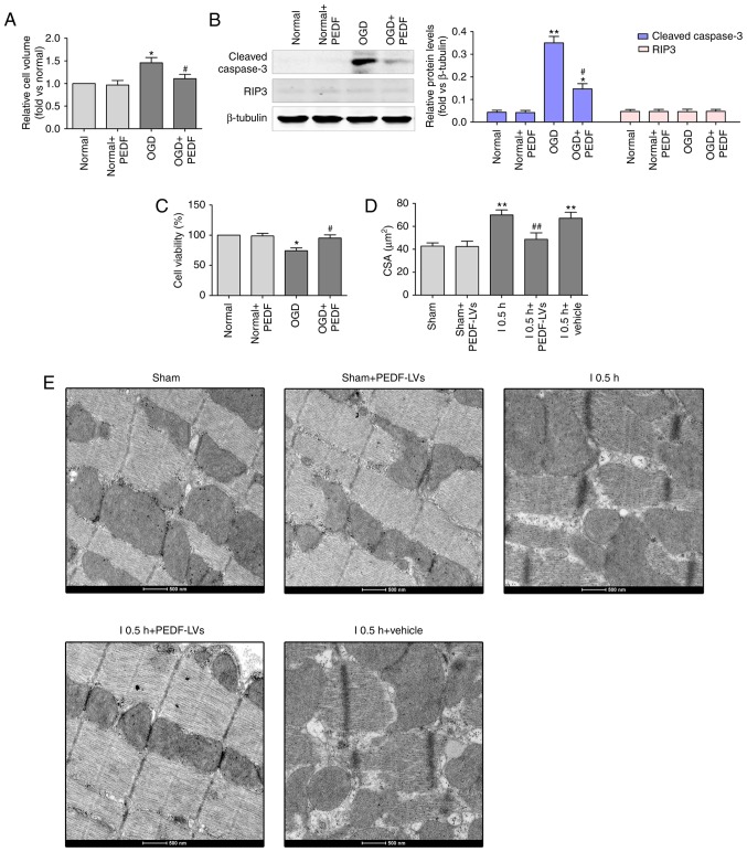
PEDF protects against OGD (ischemia)-induced cardiomyocyte edema and injury. (A) Cell volumes of 10 nmol/l PEDF-treated normal neonatal cardiomyocytes and neonatal cardiomyocytes following 30 min of OGD were measured by using confocal microscope stack scanning measurement (n=30). *P<0.05, vs. relative normal group, #P<0.05, vs. OGD group. (B) Protein levels of cleaved caspase-3 and RIP3 in PEDF-treated normal neonatal cardiomyocytes and neonatal cardiomyocytes following OGD were analyzed by western blotting (n=6). (C) A Cell Counting kit-8 assay was performed to assess cell viability in neonatal cardiomyocytes (n=6). *P<0.05 and **P<0.01, vs. relative normal controls; #P<0.05, vs. relative OGD controls. (D) Cardiomyocyte CSAs were measured following ischemia. Rats were divided into the Sham, Sham + PEDF-LVs, AMI, AMI + PEDF-LVs and AMI + vehicle groups (n=6). **P<0.01, vs. Sham group; #P<0.05 and ##P<0.01, vs. AMI group. (E) Transmission electron microscopy of cardiomyocytes (original magnification, ×11,000). OGD, oxygen-glucose deprivation; PEDF, pigment epithelium derived factor; LV, lentivirus; RIP3, receptor-interacting protein 3; AMI, acute myocardial ischemia; CSA, cross-sectional area.

