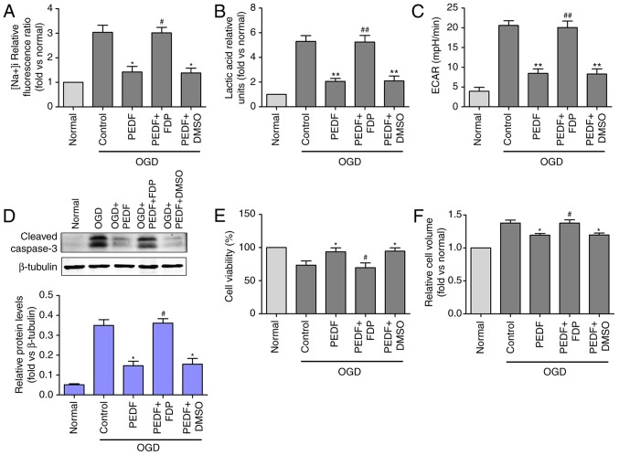
Figure 4.
Effects of PEDF on OGD-induced cardiomyocyte edema and injury are associated with decreases in glycolytic activation. (A) Na+ concentration, (B) lactic acid concentration, (C) extracellular acidification rate, (D) protein expression of cleaved caspase-3, and (E) cell viability and (F) cell volume were measured in neonatal cardiomyocytes. Cells were divided into the OGD, OGD + PEDF, OGD + PEDF + FDP, OGD + PEDF + DMSO groups (n=6). *P<0.05 and **P<0.01, vs. OGD group; #P<0.05 and ##P<0.01, vs. OGD + PEDF group. OGD, oxygen-glucose deprivation; PEDF, pigment epithelium derived factor; FDP, fructose-1, 6-diphosphate; RIP3, receptor-interacting protein 3; ECAR, extracellular acidification rate.

