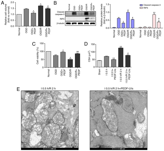
Figure 5.
PEDF inhibits cardiomyocyte edema and injury in OGD/R. (A) Cell volume, (B) protein expression of cleaved caspase-3 and RIP3, and (C) cell viability were measured in neonatal cardiomyocytes. Cells were divided into the OGD, OGD + PEDF, OGD/R and OGD/R + PEDF groups (n=6). *P<0.05 and **P<0.01, vs. OGD control group; #P<0.05 and ##P<0.01, vs. OGD/R control group. (D) CSAs in the I 0.5 h, I0.5 h + PEDF-LVs, I 0.5 h/R2 h and I 0.5 h/R 2 h + PEDF-LVs groups; the results are presented as the mean CSA of 50 random cells (n=6). **P<0.01, vs. I group; #P<0.05, vs. I 0.5 h/R 2 h group. (E) Transmission electron microscopy of cardiomyocytes (original magnification, ×11,000). OGD, oxygen-glucose deprivation; I, ischemia; R, reperfusion; PEDF, pigment epithelium derived factor; CSA, cross-sectional area; LV, lentivirus; RIP3, receptor-interacting protein 3.

