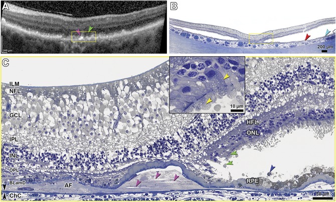
Fig. 9.

In vivo OCT and histopathology correlation of photoreceptor degeneration in GA. ILM, inner limiting membrane; GCL, ganglion cell layer; IPL, inner plexiform layer; INL, inner nuclear layer; OPL, outer plexiform layer; BLamD, basal laminar deposit; ChC, choriocapillaris. Bruch membrane, black arrowheads. A. In vivo OCT shows cRORA, OPL subsidence, inner retina thickening, and intensely hyperreflective lines (pink arrowhead) above BrM. B. Correlative histology shows atrophy of photoreceptor and RPE layers flanked by artifactual postmortem retinal detachment. Drusen, red arrowhead; SDD, teal arrowhead. C. In the yellow-framed atrophic area of Panels A and B, the ONL and RPE are absent, and avascular fibrosis (AF) is seen beneath BLamD. Clefts in the AF (pink arrowheads) correlate to the hyperreflective lines in panel A. Distances from the top-bottom surfaces of the clefts (pink arrowheads) to the inner collagenous layer of BrM are (from left to right) 11.8 to 10.2 µm, 30.4 to 26.8 µm, and 23.8 to 19.9 µm, respectively. In the nonatrophic area, near the descent of ELM (green arrowheads), photoreceptor nuclei and mitochondria (yellow arrowheads, inset) are retracted toward HFL. Sloughed RPE, blue arrowhead. At the upper right is artifactual thickening of inner retina due to edema (See Figure S1, Supplemental Digital Content 1, http://links.lww.com/IAE/A954).
