Figure 4.
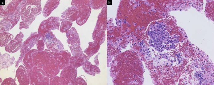
Scanned low magnification (a, ×5) and high magnification (b, ×20) images of hematoxylin-and-eosin–stained slide from the primary GBC specimen obtained through EUS-FNA demonstrating small amount of adenocarcinoma. EUS-FNA, endoscopic ultrasound-guided fine needle aspiration; GBC, gallbladder carcinoma.

