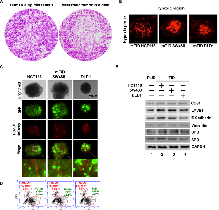
Fig. 2.
(A) The architecture of mTiD cultures resemble that of metastatic tumor in the lung of patients. H&E stained sections of metastatic colon tumor in the lung compared to a mTiD organoid demonstrates micrometastatic tumor deposits. (B) mTiD cultures have a hypoxic core. A) mTiD-HCT116, B) mTiD-SW480 and C) mTiD-DLD1 cultures were stained with Hypoxyprobe-Red549. Hypoxic regions (red) were observed in the core of the organoids. (C) GFP labeled HCT116, SW480 or DLD1 colon cancer cells were grown in presence of PLiD consisting of NL20, MrC5, HLEC and mCherry labeled HUVEC cells. Fluorescence imaging demonstrates distribution of colon cancer (green) and HUVECs (red) in mTiD organoids. Merged images show blood vessel-like tubules of red HUVEC cells in the mTiD. (D) Flow cytometry shows the percentage of GFP labeled colon cancer and m-Cherry labeled HUVEC- cells mTiD cultures. (E) Western blot analyses demonstrate the expression of CD31, LYVE1, E-cadherin, vimentin, SPD and SPB in PLiD (devoid of cancer cells) and mTiD cultures.

