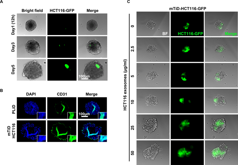
Fig. 3.
Colonization and angiogenesis in mTiD organoids. (A) Live cell imaging shows colonization of HCT116-GFP cells in PLiD live culture in time dependent manner. (B) Immunocytochemical staining shows CD31 positive staining appears like single tube-like formation in PLiD section and mTiD sections. (C) Pretreatment of PLiD with increasing concentrations of cancer cell derived exosomes enhances colonization by cancer cells.

