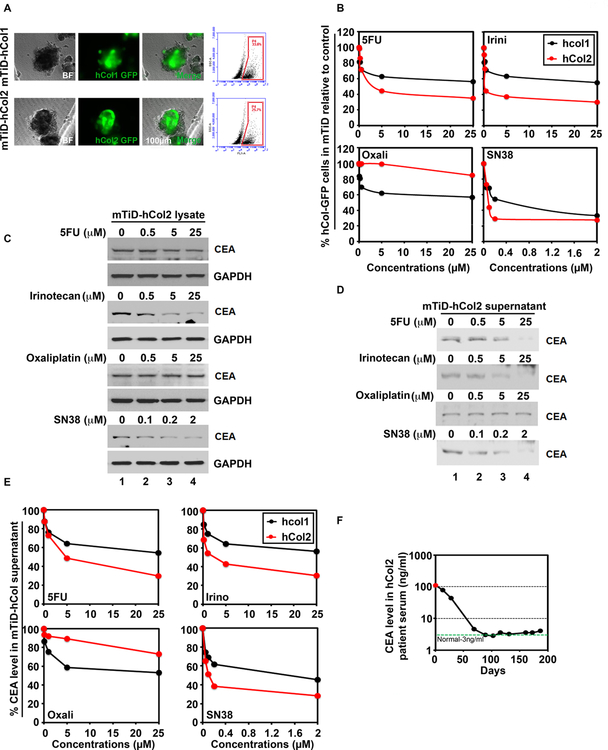
Fig 6.
mTiD is an effective platform to screen drug responses in primary patient-derived colon cancer cells. (A) Representative bright field (BF), GFP-positive cells and merged images of mTiD generated with GFP-labeled primary colon cancer cells isolated from 2 patients (hCol1 and hCol2) are shown. Enzymatically dissociated primary colon mTiD were flow sorted for GFP-positive cells. The mTiD cultures contained 25.7 to 33.8% tumor. (B) Dose-response curves were generated for GFP-labeled primary colon cancer cells in mTiD cultures, after 48h treatment with 5FU, oxaliplatin, irinotecan or SN38 followed by flow sorting. (C) Reduction in carcinoembryonic antigen (CEA) levels in primary colon mTiD cultures corresponds to decrease in serum CEA from the same patient. The hCol1 mTiD cultures were assessed for CEA levels using western blotting of cell lysates and, (D) cell supernatants after treatment with varying doses of 5FU, oxaliplatin (Oxa), irinotecan (Iri), or SN38. (E) Chemiluminescence immunoassay (CLIA) of supernatants from mTiD-hCol2 cultures demonstrates a decrease in CEA levels upon 5FU, Iri and SN38 treatments but not oxaliplatin. (F) CEA levels were measured by CLIA in hCol2 patient serum after chemotherapeutic (FOLFIRI) treatment. Data shows significant linear decrease in CEA serum levels after FOLFIRI treatment.

