Fig 7.
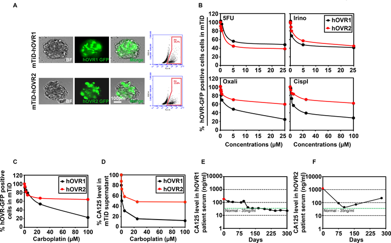
mTiD is an effective platform to screen drug responses in primary patient-derived ovarian cancer cells. (A) Representative bright field (BF), GFP-positive cells and merged images of mTiD generated with GFP-labeled primary ovarian cancer cells isolated from 2 patients (hOVR1 and hOVR2) are shown. Enzymatically dissociated primary colon mTiD were flow sorted for GFP-positive cells. The mTiD cultures contained 23 to 25.7% tumor. (B) Dose-response curves were generated for GFP-labeled primary ovarian cancer cells in mTiD cultures, after 72 h treatment with 5FU, oxaliplatin, irinotecan or cisplatin followed by flow sorting. 5FU and irinotecan reduced the number of GFP positive hOVR1 and 2 cells. Cisplatin and oxaliplatin reduced the number of hOVR1 cells but not hOVR2 by over 50%. (C) mTiD cultures of GFP-labeled hOVR cells were treated with carboplatin for 72h followed by flow cytometry. hOVR1 (black line) were more sensitive to carboplatin at increasing concentration than hOVR2 cells (red line). (D) Chemiluminescence immunoassay (CLIA) for CA125 in supernatants from mTiD-hOVR1 demonstrated a greater reduction in CA125 levels than mTiD-hOVR2 cultures upon carboplatin treatment. (E) CA125 levels are reduced in the serum from hOVR1 patient responsive to carboplatin treatment. (F) CA125 levels in carboplatin refractory patient hOVR2 showed higher levels of CA125 in the serum.

