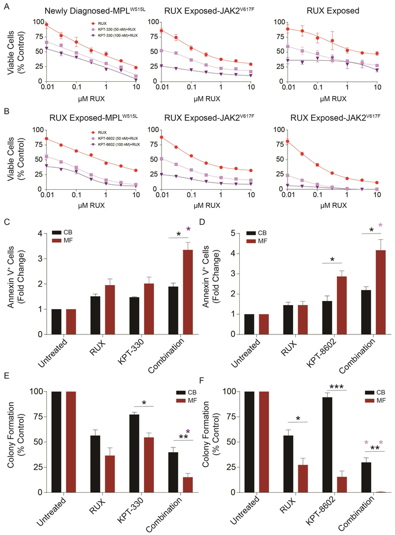
Figure 5. Inhibition of NCT by KPT-330 or KPT-8602 enhances effect of ruxolitinib on primary MF CD34+ cells.
Primary MF CD34+ cells were treated with either ruxolitinib alone or in combination with a fixed concentration of KPT-330 at 50 or 100 nM (A) or KPT-8602 at 50 or 100 nM (B) for 72 h. Viable cells were quantified by MTS assay. Each plot represents one sample from one patient. When known, genotypes and treatment status are shown. Inhibition of NCT by KPT-330 at 50 nM (C) or KPT-8602 at 50 nM (D) increased ruxolitinib (100 nM)-induced apoptosis (n=3 each, 72 h). Primary MF or CB CD34+ cells were treated with indicated concentrations of either ruxolitinib alone or in combination with KPT-330 or KPT-8602 for 72 h, and apoptosis was measured with Annexin V. KPT-330 at 100 nM (E) or KPT-8602 at 50 nM (F) enhanced ruxolitinib (1.5 µM)-induced inhibition in colony formation of primary MF vs. CB CD34+ cells (n=3–7). Statistical significance was compared between MF vs. CB (*p<0.05; **p<0.01). Purple asterisks represent combination groups that are statistically distinct (p<0.002 for each) from the ruxolitinib group.

