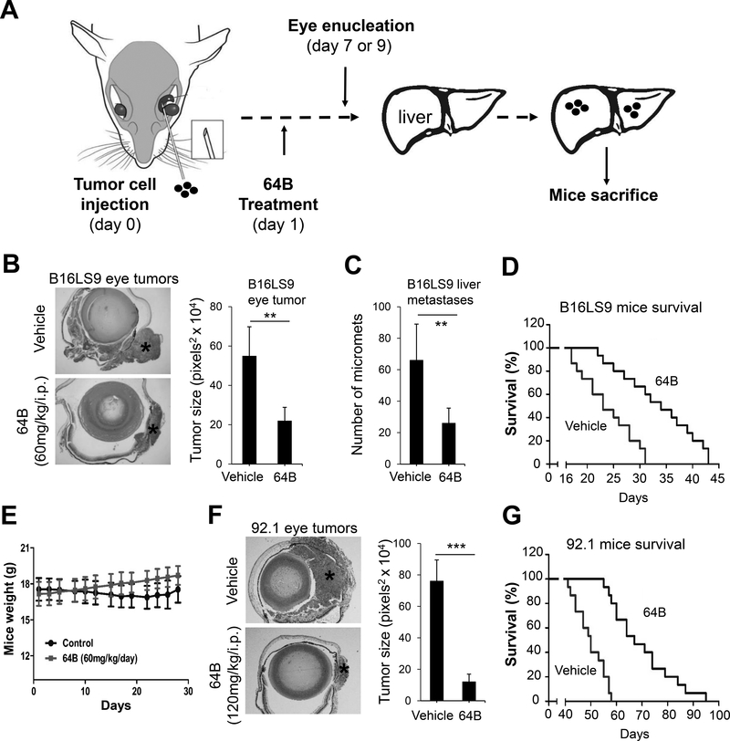
Figure 5.
64B inhibits primary eye tumor growth, liver metastasis, and prolongs survival in UM mouse models. A, Schematic showing timeline and procedure for the animal experiments. B, C, D, E, B16LS9 mouse skin melanoma cells (2 × 10⁶/ eye) were injected in the sub-uveal area of C57BL/6 mice eyes where they form melanomas in the uvea. One day after eye inoculation (day 1), mice (8/group) were treated with either vehicle control or 60mg/kg/i.p of 64B 5 days per week. On day 7, tumor-bearing eyes were enucleated, fixed and stained to evaluate tumor burden (B). Representative H&E sections are shown (Left) and quantification of eye tumor (*) size was performed in 4 randomly selected mice/group (Right). Mice were sacrificed on day 28 and number of liver metastases counted (8 mice/group) (C). In a repeat experiment with identical design, mice were kept under treatment after eye enucleation and terminated when reaching IACUC criteria to measure survival by Kaplan-Meier analysis (15 mice/group) (D). E. Weight of mice used in (D). F, G 92.1 human uveal melanoma cells (2 × 10⁶/ mouse) were injected in the suprachoroidal space of 3 ½ month old female nu/nu mice (Envigo Corp; Strain: Hsd: Athymic Nude-Foxn1nu) (15 mice/group). On day 1, mice were treated with either vehicle control or 120mg/kg/i.p of 64B 5 days per week. On day 7, eyes were removed and evaluated for tumor burden. Representative H&E stained eyes are shown (left) and tumor size (*) quantified in 6 mice per group (F). Survival rate of mice (15 mice/group) (G).

