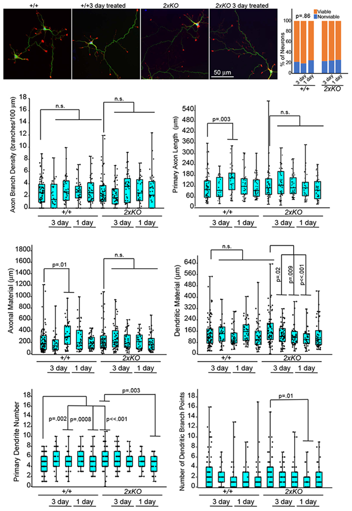
Figure 4:

Effects of Patient CSF on neuronal viability and development. E15.5 cortical neurons from wildtype (+/+) and Trim9−/−:Trim67−/− (double KO) mice were treated with patient CSF (1:100) for 3 days or 1 day prior to fixation. Neurons were stained for βIII tubulin (green), filamentous actin (red), and DNA (blue). Several parameters were measured per neuron, including density of axon branches, primary axon length, total axonal material, dendritic material, primary dendrite number, and dendrite branch points. In the box and whisker plots: the cyan boxes for each genotype are untreated. The second two were incubated from the 24th to the 96th hour (3 days) with 1:100 CSF from patient 1 (magenta boxes) and patient 2 (yellow boxes), respectively, the next two were incubated from the 72nd to the 96th hour (1 day), patient 1 and patient 2, respectively. Boxplots depict median +/− the interquartile range (IQR), whiskers reach minimum and maximum values. P-values were determined by ANOVA, comparisons made to untreated control, with Bonnferroni post hoc corrections of the number of comparisons made. P- values are considered significant when p<0.05. No consistent significant effects of patients’ CSF on neurons were observed.
