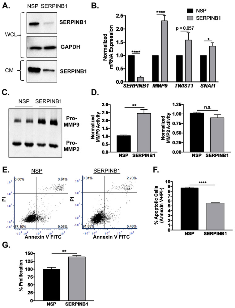
Figure 3. SERPINB1 loss stimulates EMT and a proliferative phenotype in normal prostate cells.
A. RWPE-1 cells were transfected with non-specific (NSP) and SERPINB1-specific siRNA, and knockdown was verified using Western blot. GAPDH was used as a loading control. A representative blot is shown. WCL=whole cell lystate, CM=complete media (secreted SERPBINB1). B. mRNA expression of SERPINB1 and EMT markers MMP9, TWIST1, SNAI1 were determined in RWPE-1 cells after SERPINB1 knockdown using quantitative PCR and normalized to GAPDH. Data were normalized to NSP treated samples, and differences were assessed using Student’s t-test (n = 7; * p < 0.05, **** p < 0.0001). C. Expression and activity of MMP2 and MMP9 were determined in RWPE-1 cells after SERPINB1 knockdown using gelatin zymography. A representative gel is shown. D. Band densitometry of MMP9 and MMP2 was performed using ImageJ, and normalized differences were assessed using Student’s t-test (n = 3; ** p < 0.01, n.s. = not significant). E. Apoptosis was assessed in RWPE-1 cells after transfection with NSP and SERPINB1 siRNA via PI and Annexin V staining. Representative plots are shown. F. Difference in apoptosis (% Annexin V+/PI− cells) was determined using Student’s t-test (n = 3; **** p < 0.0001). G. Proliferation was assessed in RWPE-1 cells after transfection with NSP and SERPINB1 siRNA via colorimetric BrdU incorporation assay. Data were normalized to NSP treated samples, and difference was assessed using Student’s t-test (n = 3; ** p < 0.01).

