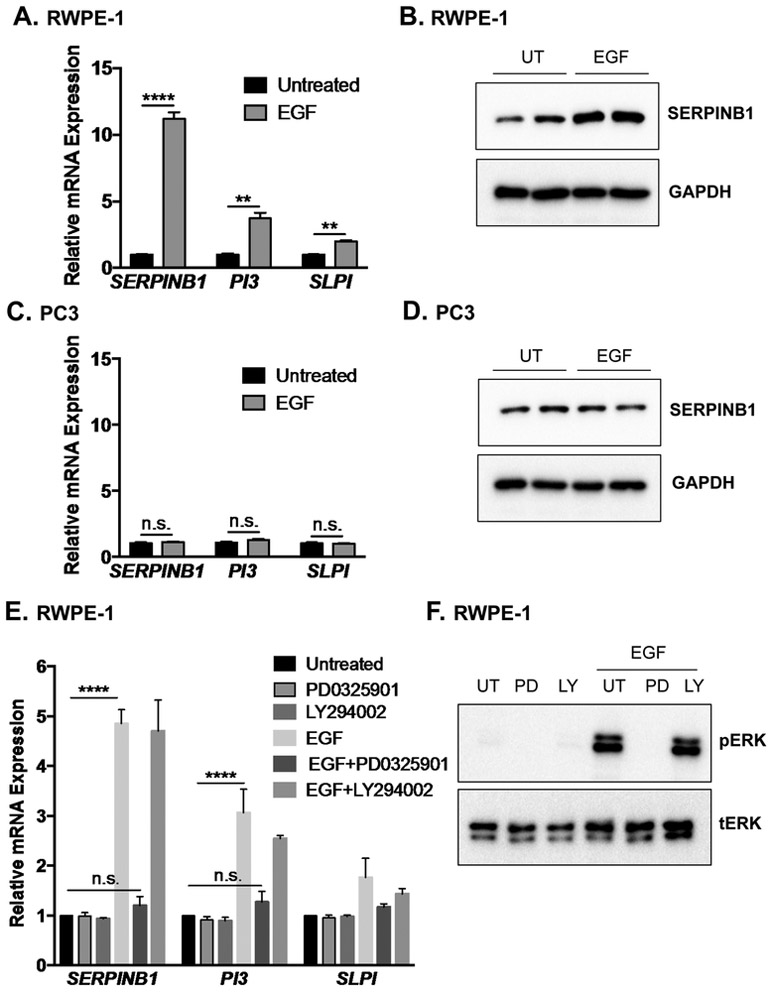
Figure 4. SERPINB1 expression is induced via ERK1/2 signaling in normal prostate but not prostate cancer cells.
A. RWPE-1 cells were serum starved and treated with epidermal growth factor (EGF; 20 ng/mL) for 4 hours. mRNA expression of SERPINB1, PI3, and SLPI were determined using quantitative PCR and normalized to GAPDH. Data were normalized to untreated samples, and differences were assessed using Student’s t-test (n = 3; ** p < 0.01, *** p < 0.001). B. RWPE-1 cells were serum starved and left untreated (UT) or treated with EGF (20 ng/mL) for 24 hours. SERPINB1 expression was determined using Western blot. GAPDH was used as a loading control. A representative blot with two of each treatment condition is shown. C. PC3 cells were serum starved and left untreated or treated with EGF (20 ng/mL) for 4 hours. mRNA expression of SERPINB1, PI3, and SLPI were determined using quantitative PCR and normalized to GAPDH. Data were normalized to untreated samples, and differences were assessed using Student’s t-test (n = 3, n.s. = not significant). D. PC3 cells were serum starved and treated with EGF (20 ng/mL) for 24 hours. SERPINB1 expression was determined using Western blot. GAPDH was used as a loading control. A representative blot with two of each treatment is shown. E. RWPE-1 cells were serum starved and treated with EGF (20 ng/mL) in the presence of MEK inhibitor PD0325901 (0.25 μM) or PI3K inhibitor LY294002 (0.25 μM) for 4 hours. mRNA expression of SERPINB1, PI3, and SLPI were determined using quantitative PCR and normalized to GAPDH. Data were normalized to untreated samples, and differences were assessed using ANOVA with post-hoc Tukey (**** p < 0.0001, n.s. = not significant). F. RWPE-1 cells were serum starved and treated with EGF (20 ng/mL) in the presence of PD0325901 (0.25 μM) or LY294002 (0.25 μM) for 30 minutes. pERK1/2 and tERK1/2 levels were examined via Western blot.

