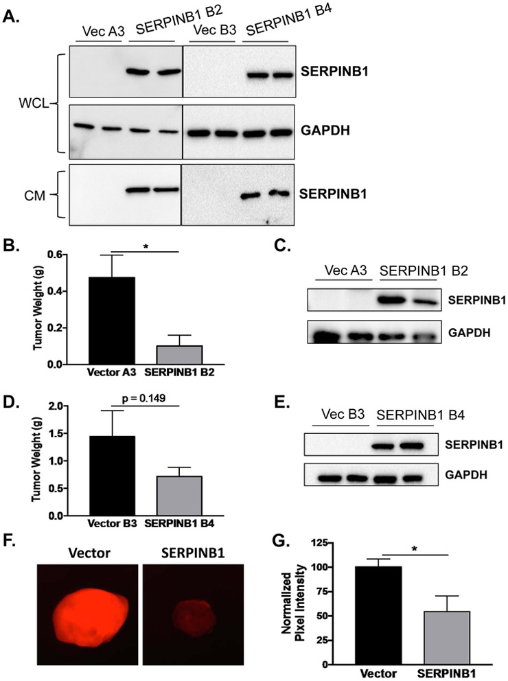
Figure 6. SERPINB1 overexpression reduces prostate cancer xenograft growth in vivo.
A. SERPINB1 was detected in whole cell lysates (WCL) and concentrated conditioned media (CM) collected over 24 hours in C4-2 cells stably expressing SERPINB1 (SERPINB1 clones B2 and B4) and vector controls (Vector clones A3 and B3). GAPDH was used as a loading control. Representative blots are shown. B. Tumor weight was compared between Vector A3 and SERPINB1 B2 xenografts injected subcutaneously into the flanks of athymic nude mice using Student’s t-test (n = 10 for Vector, n = 3 for SERPINB1; * p < 0.05). C. SERPINB1 was detected in harvested tumors at the end of the experiment using Western blot. A representative blot is shown. D. Tumor weight was compared between Vector B3 and SERPINB1 B4 xenografts injected subcutaneously into the flanks of athymic nude mice using Student’s t-test (n = 10 for Vector, n = 11 for SERPINB1; p = 0.149). SERPINB1 was detected in harvested tumors at the end of the experiment using Western blot. A representative blot is shown. F. Intra-tumoral neutrophil elastase activity was measured ex vivo using an NE specific optical probe. G. Intra-tumoral NE activity was quantified using ImageJ and normalized to Vector controls. Difference was assessed using Student’s t-test (n = 8 for Vector, n = 4 for SERPINB1; * p < 0.05).

