
Figure 2. Atherosclerotic pathology in aorta.
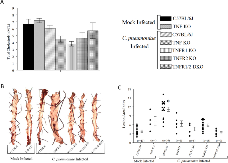
Groups of 6–8-week-old male WT, TNF KO, TNFR1 KO, TNFR2 KO, or TNFR 1/2 DKO were infected on day 0, 15, and 30 with CPN. Separate groups of WT and TNF KO mice were infected with SPG buffer (mock). Mice were bled and euthanized on day 105 after primary inoculation for serum cholesterol assay and pathology estimation. A, Mean ± SEM of serum cholesterol levels in each group is shown. B, Representative images of qualitative pathology estimation using oil red O staining. The pins used to secure tissues to the gel are also seen in the pictures. C, Quantitative estimation of atherosclerotic pathology. Each marker represents an individual aorta/animal. Mean ± SEM for each group are also shown. The number of animals within each group are shown on the X-axis. * Significant (p≤0.05; ANOVA) between CPN-infected WT and all other groups. Results are pooled from two independent experiments.