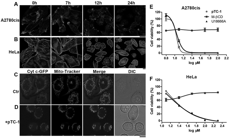
Figure 2.
pTC-1 interacts with cell cytoskeleton and induces mitochondria fission. After incubating pTC-1 with (A) A2780cis or (B) HeLa cells to reach designated time points, we used Alexa Fluor 633 phalloidin (F-actin) to reveal the changes of actin filaments. The concentration of pTC-1 is 12.5 µM for A2780cis cell and 25 µM for HeLa cell. (C) and (D) pTC-1 (25 µM) induced fragmental mitochondria in 2H18 HeLa cells (expressing GFP labeled Cyt c). After treated 2H18 HeLa cells for 22 h by pTC-1, we also used Mito-Tracker to stain cellular mitochondria. 48 h cell viability of (E) A2780cis or (F) HeLa without or with addition of M-βCD and U18666A. The concentrations of M-βCD for treating A2780cis and HeLa cells are 5 and 2 mM, respectively. The concentration of U18666A for treating A2780cis and HeLa cell is 1 µg/mL. Scale bar in (A) to (D) is 10 µm.

