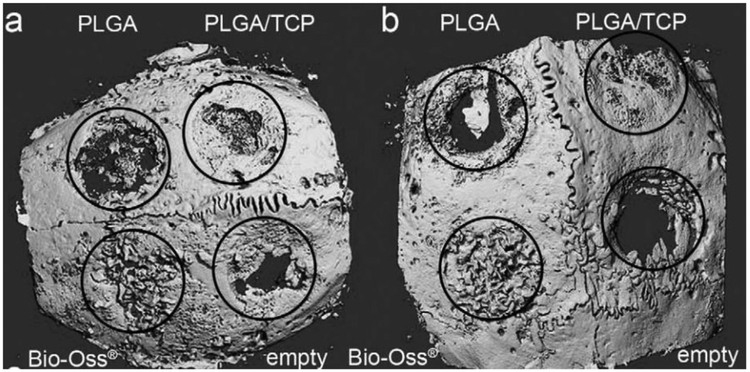
Figure 11.
Micro-computed tomography of the cranial defects (diameter=6 mm) in New Zealand White rabbits after 4-week implantation using PLGA, PLGA/TCP composites. (a, b) Two examples of the CT of the entire cranial bone are shown. Defect margins and treatment modalities are indicated. Adapted from Ref [92].

