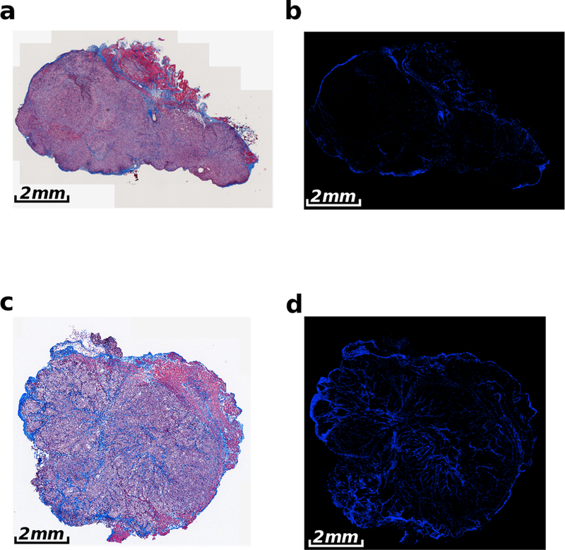
Figure 2:
Representative Masson’s trichrome stained images of AsPC-1 and BxPC-3 tumors implanted into the pancreas of nude mice. Showing (a) AsPC-1 tumor and (b) color-segmented collagen distribution of the AsPC-1 tumor. (c) BxPC-3 tumor with (d) corresponding color-segmented collagen distribution. The AsPC-1 tumors in (a) and (b) have a collagen density of 5.6 ± 1.4%, in comparison to the collagen density of 12.5 ± 1.1% for the BxPC-3 tumor in (c) and (d).

