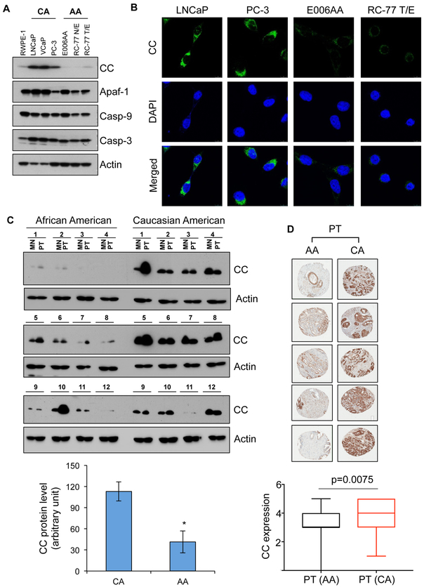
Figure 1. CC, a key component of apoptosome and OXPHOS system, is reduced in PCa cell lines and tumor specimens derived from AA men with PCa.
A, Expression of the components of apoptosome complex, which include CC, Apaf-1, caspase-9 (Casp-9), and caspase-3 (Casp-3) were examined using immunoblotting in RWPE-1 (normal prostate epithelial cells), LNCaP, VCaP, PC-3, E006AA, RC-77 N/E and RC-77 T/E cells. Actin serves as a loading control. B, Expression of CC in LNCaP, PC-3, E006AA and RC-77 T/E cells using immunofluorescence. C, Immunoblot analysis of CC in primary tumor (PT) and matched non-tumor (MN) prostatic tissues from AA and CA men with PCa. Actin serves as a loading control. Densitometry analysis of immunoblots of CC in PT tissues from AA and CA men with PCa (n=12 for each race). D, Immunohistochemistry (IHC) analysis of CC expression in PT in AA and CA men with PCa using tissue microarray (TMA) sections. Scoring analysis of IHC of CC in PT tissues from AA (n=92) and CA (n=89) men with PCa. Data in C represent mean ± SD of n=12. Significant differences between means were assessed using analysis of variance (ANOVA) and GraphPad Prism Version 6.0. *p < 0.05 vs CA primary tumor.

