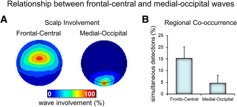
Figure 3.

Relationship between frontal-central and medial occipital delta waves. A, Typical scalp involvement for the two types of delta waves. For each delta wave detected in the two electrodes of interest (frontal-central and medial-occipital), the number of concurrent detections (in a window of 200 ms, centered on the wave negative peak) in other electrodes was computed. Therefore, each map shows the number of cases in which each electrode showed a co-occurring delta wave (the electrode of interest has 100% value in each map). B, Relative occurrence of simultaneous detections (of the same type/duration) across the two electrodes of interest.
