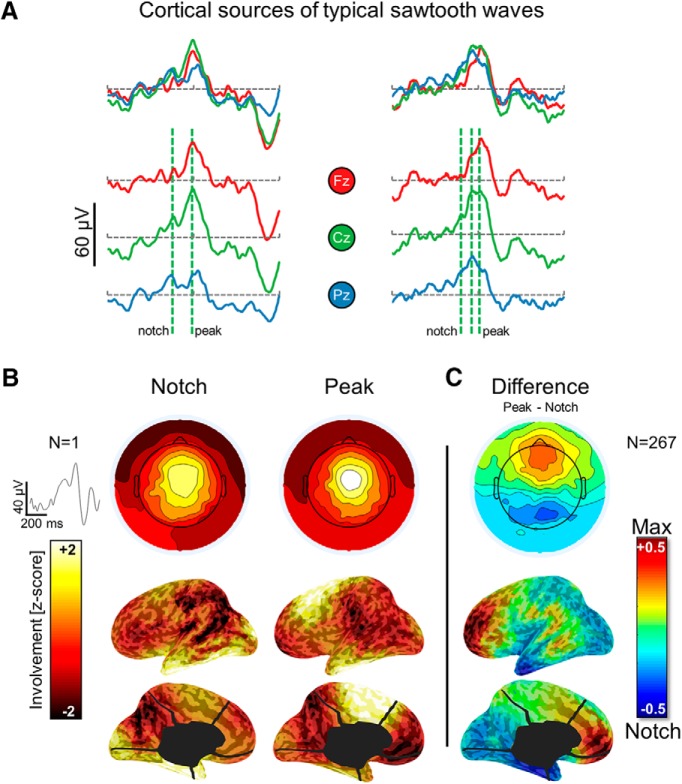
Figure 6.
Cortical sources of typical sawtooth waves. A, EEG traces (three electrodes, corresponding to Fz, Cz, and Pz; negative-up on the y-scale) of two representative notched sawtooth waves (±250 ms around the negative peak of the algorithm detection). B, Scalp and cortical involvement corresponding to the notch and the maximal negative peak of a single sawtooth wave. C, Difference in average involvement between the notch and maximal negative peak (computed across 267 sawtooth waves in one representative subject; similar results were obtained in other participants). For this evaluation, all detections in one subject were visually inspected to identify typical sawtooth waves with a negative amplitude >20 μV and a well recognizable notched, triangular shape. The timing of the notch and the maximal negative peak were marked manually. Differences shown in C are statistically significant over all voxels (p < 0.05, corrected).

