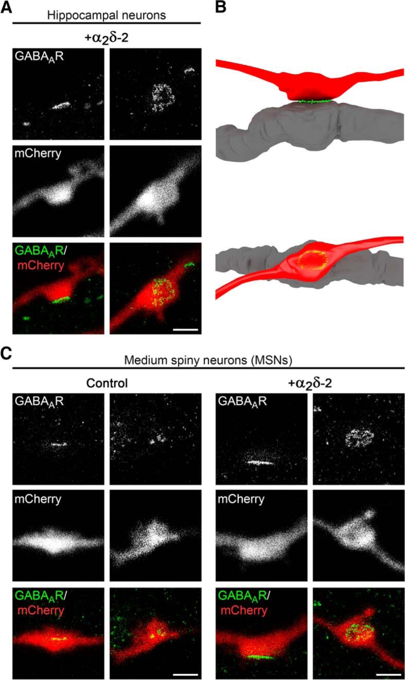
Figure 3.

GABAAR clusters are confined to the postsynaptic membrane. gSTED micrographs of cultured hippocampal (A) or MSNs (C) transfected with α2δ-2 and mCherry or mCherry only (control; 20–30 DIV). Transfected neurons (red, detected in confocal mode) were immunolabeled for the GABAAR (green, detected in gSTED mode). In all conditions, postsynaptic GABAAR clusters are closely opposed to mCherry-positive presynaptic boutons. B, 3D model showing that the GABAAR staining pattern depends on the orientation of the imaged synapse, which applies both for hippocampal as well as MSNs. Scale bar, 1 μm.
