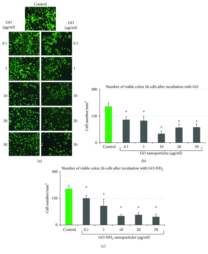
Figure 4.
Quantitative evaluation of cellular viability of Colon 26 cells after incubation for 24 hours with pristine and aminated GO. (a) Fluorescent micrographs of FDA-stained Colon 26 cells incubated for 24 hours in the presence of GO and GO-NH2 nanoparticles at different concentrations. Magnification 10x; bar 100 μm. (b) Number of viable Colon 26 cells after 24 hours of incubation in the presence of GO nanoparticles. (c) Number of viable Colon 26 cells after 24 hours of incubation in the presence of GO-NH2 nanoparticles (asterisks ∗ denote p < 0.05, respectively, when the tested probes are compared to untreated cells).

