Figure 7.
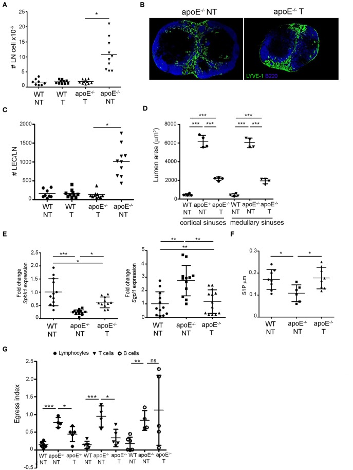
Reversing dyslipidemia improves lymphocyte egress and LN hypertrophy. (A) WT and apoE−/− mice were treated with eztimibe (treated, T) or vehicle (non-treated, NT) and LN cellularity expressed as fold change over WT mice was determined. n = 8–10 mice per group; *p < 0.05. (B) Immunoreactivity for B220 and LYVE-1 was examined in LN sections from NT and T apoE−/− mice. Images are representative of three independent experiments (n = 3–5). (C) The number of LECs was determined in LN suspensions from NT WT, NT and T apoE−/− mice by flow cytometry and expressed as fold change over baseline. n = 8–10; *p < 0.05. (D) Lumen area of cortical and medullary sinuses was determined on LN sections NT WT, NT, and T apoE−/− mice. n = 4 mice per group; ***p < 0.0005. (E) Quantitative RT-PCR analysis was performed on whole LN RNA to examine expression of Sph1k and Spgl1 in NT WT, NT, and T apoE−/− mice. Data is pooled from three independent experiments with 4 mice per group in each experiment; *p < 0.05; **p < 0.005; ***p < 0.0005. (F) S1P content was analyzed in efferent lymph of NT WT, NT and T apoE−/− mice by mass spectrometry. Data is pooled from three independent experiments with 3–4 mice per group in each experiment (G) Egress of adoptively transferred CD45.1 lymphocytes, T and B cell from NT WT, NT and T apoE−/− mice was examined. n = 4–6 mice per group; *p < 0.05; **p < 0.005; ***p < 0.0005.

