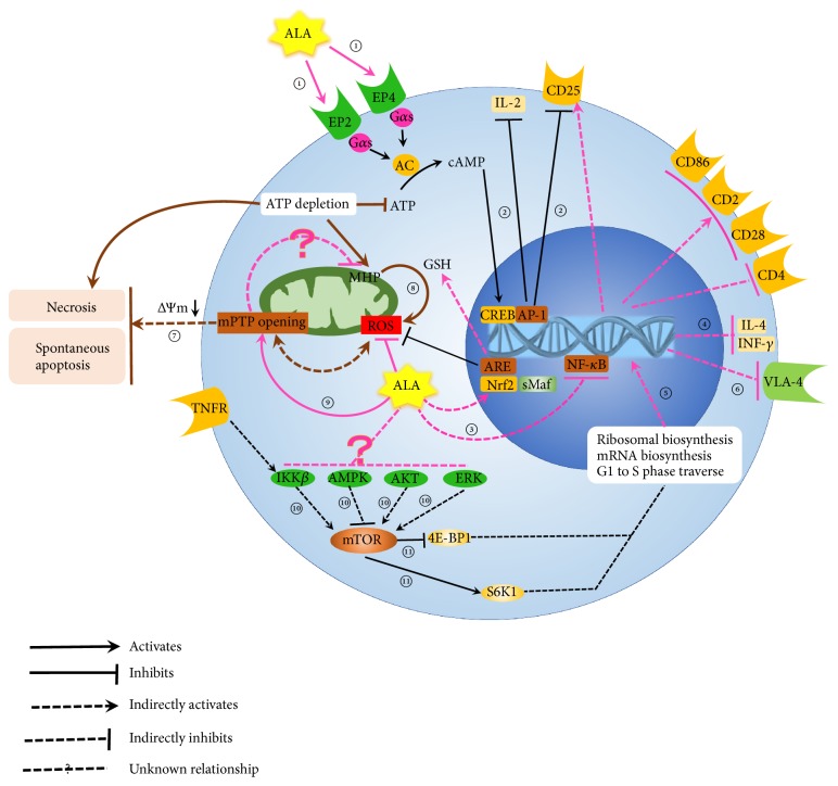
Figure 1.
Effect of ALA on T cell. ① ALA increase cAMP synthesis through activation of prostaglandin receptors (EP2 and EP4) in peripheral blood T cells. ② The expression of IL-2 and IL-2Rα (CD25) could be inhibited when the level of cAMP was increased by ALA. ③ ALA could inhibit NF-κB activation induced by TNF in Jurkat T cells. ④ ALA could suppress production of interferon-γ (IFN-γ) and interleukin -4 (IL-4). ⑤ ALA could induce lymphocyte progression from G0/G1 to S phase. ⑥ ALA was also reported to reduce migration of T cell, lymphocyte and monocyte of models of MS, and Jurkat T cells, which were associated with down-regulated expression of very late activation-4 antigen (VLA-4). ⑦ Uncontrolled mPTP opening leads to decrease ∆Ψm irreversibly until dissipation which results in apoptosis and necrosis of cells. ⑧ Persistent MHP in SLE T cells could enhance ROS production. ⑨ Studies showed that ALA and DHLA promoted mPTP opening in mitochondria of rat liver. ⑩ mTORC1 was a central common regulator of a complex signaling network In cytoplasm, in which Ras/Erk, PI3K/Akt, and IKKβ activated mTORC1 while Dsh/GSK3 and LKBl/AMPK inactivated mTORC1. ⑪ Activated mTORC promoted protein synthesis by phosphorylating the eukaryotic initiation factor 4E-binding protein 1 (4E-BP1) and p70 ribosomal S6 kinase 1 (S6K1).

