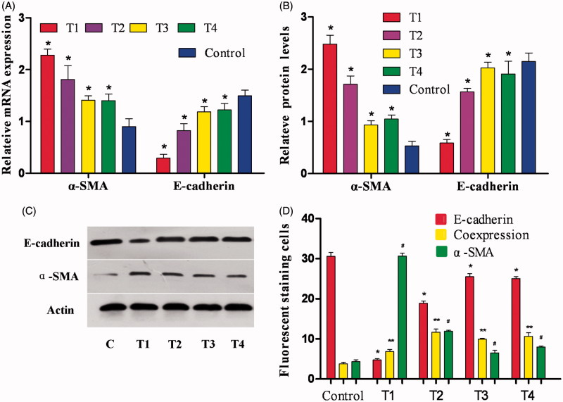
Figure 2.
Effect of fasudil on HK-2 cells. (A) HK-2 cells were cultured in each group as discussed in the Methods section. RT-PCR was performed and the expression of E-cadherin and α-SMA was normalized to actin. Results are from three independent experiments performed in triplicate and are displayed as the relative expression normalized to the control samples. Values are the means with S.E.M. shown by vertical bars. *p < .05. (B) Densitometric analysis for western blots. Values are the means from triplicate determinations with the S.E.M. shown by vertical bars. *p < .05. (C) Western blot for E-cadherin and α-SMA using HK-2 cells treated as aforementioned. (D) The number of positive-stained cells by immunofluorescence is shown by vertical bars. *, ** and #p < .05.

