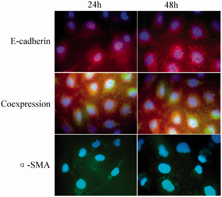
Figure 3.
Effect of fasudil on cell immunofluorescence. Immunofluorescence staining of HK-2 cells captured by inverted fluorescence microscope at 100× magnification. The number of cells from 10 random fields for each group was calculated. Data are presented as the mean ± S.E.M. *p < .05 versus control. Red: E-cadherin expression; Green: α-SMA expression; Blue: nuclei (DAPI). Cells coexpressing α-SMA and E-cadherin are yellow. Results are from three experiments performed in triplicate.

