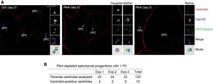
Identification of the daughter centriole through Centrobin. Ependymal progenitors treated as in Fig
4B were fixed at day 0 and immunostained for Centrobin and Cep152. GFP‐Centrin1 served as both infection and centriole markers. Zo‐1 was visualized in the same channel with Centrobin to label the cell boundaries because their antibodies were from mouse and their staining patterns did not overlap. Parental centrioles are denoted by arrows. An illustration is provided for each set of the magnified (2×) images. Scale bar, 1 μm.

