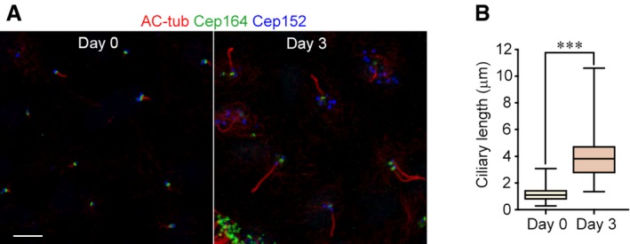
Figure EV3. Ciliary length increases dramatically during the differentiation of ependymal progenitors.

- Cilia in the progenitors (day 0) and mEPCs (day 3). Ependymal progenitors treated as in Fig 4B were fixed and immunostained to visualize acetylated tubulin (AC‐tub; a cilia marker), Cep164 (a marker for mother centriole or basal body), and Cep152 (a marker for parental centriole and deuterosome). Scale bar, 5 μm.
- Quantification for the ciliary length. The results were from three independent experiments. At least 105 cilia were measured in each experiment and condition. The bottom and top of the box represent the 25th and 75th percentiles, respectively. The band is the median. The ends of the whiskers indicate the maximum and minimum of the data. Two‐tailed unpaired Student's t‐test: ***P < 0.001.
