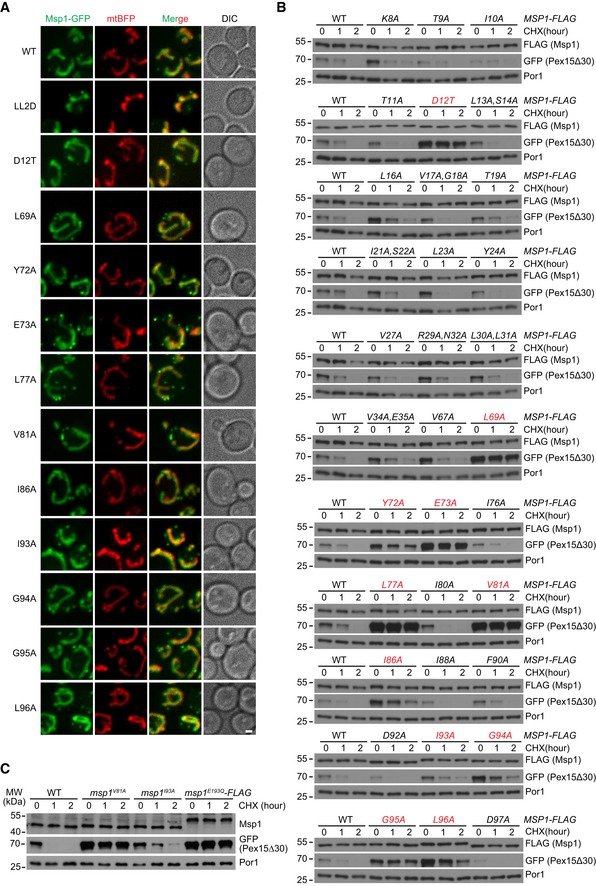
The normal localization of Msp1 N‐domain mutants. Z projections and DIC images are shown. Mitochondrial Msp1‐GFP colocalizes with mtBFP. Peroxisomal Msp1 appears as extra‐mitochondrial dots. Scale bar represents 1 μm.
Degradation of GFP‐Pex15Δ30 in WT and Msp1 N‐domain mutant cells. Mutants displaying defective degradation are highlighted in red.
Degradation of GFP‐Pex15Δ30 in untagged Msp1 N‐domain mutant cells. msp1
E193Q
‐FLAG strain is used as a control for Msp1 loss of function.
Data information: In this figure, WT and mutant forms of Msp1‐GFP and Msp1‐FLAG were expressed from the endogenous chromosomal locus, and GFP‐Pex15Δ30 was expressed from a centromeric plasmid under the control of
TEF1 promoter.

