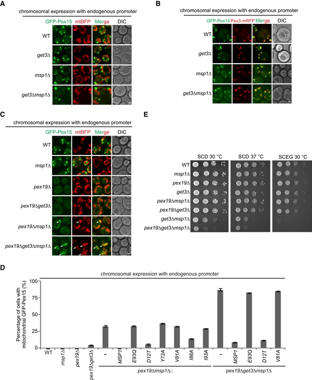
Figure 5. Pex15 is mislocalized to mitochondria and removed by Msp1 in pex19∆ cells.

- Chromosomally expressed GFP‐Pex15 driven by endogenous promoter did not mislocalize to mitochondria in GET3 and MSP1 mutant strains.
- GFP‐Pex15 expressed as in (A) localizes to peroxisome in GET and MSP1 mutant strains. Pex3 was chromosomally tagged with mRFP to visualize peroxisomes.
- Localization of GFP‐Pex15 expressed as in (A) in the indicated stains. Arrows point to mislocalized GFP‐Pex15 on mitochondria.
- The percentage of cells with mitochondrial GFP‐Pex15 in the indicated strains. Data values represent means ± SEM from three independent experiments, with at least 100 cells counted in each experiment.
- The indicated strains were grown in glucose media to log phase and then spotted on SCD or SCEG plates in a 10‐fold serial dilution, and then incubated at indicated temperatures for 2–5 days.
