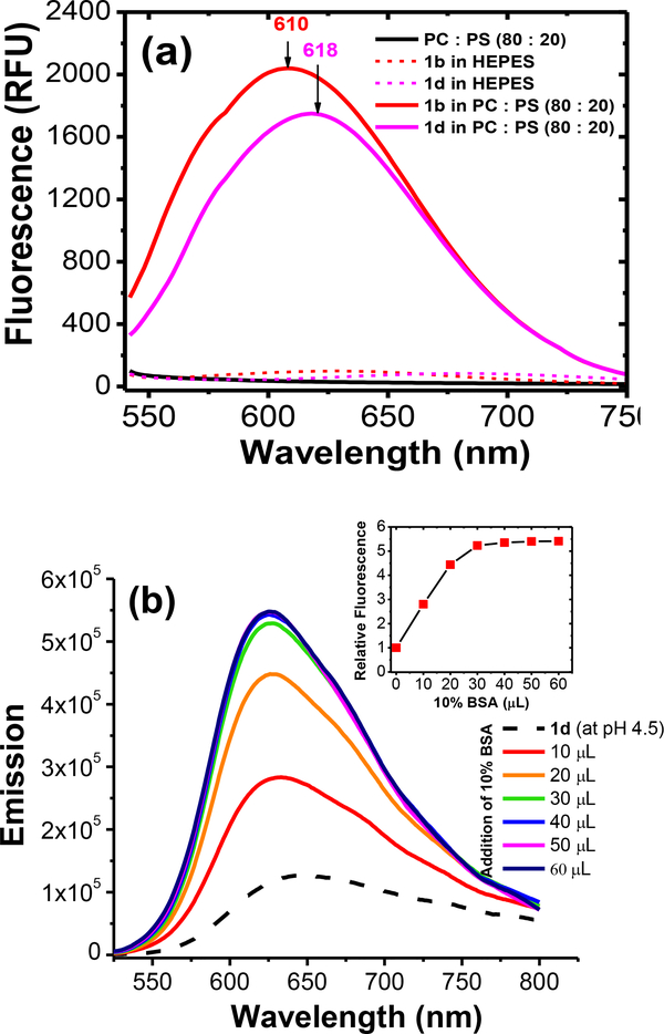
Figure 4.
(a) Fluorescence emission obtained for 2b and 2d (1 × 10⁻⁵ M) upon introduction in to 1 mM lipid vesicles (PC:PE 80:20) in HEPES buffer (pH = 7.4). Probes were excited at 530 nm. (b) Represents the fluorescence emission enhancement observed in 2d (1 × 10⁻⁵ M) upon addition of 10% bovine serum albumin (BSA) in to an aqueous solution (pH 4.5) of probe 2d (1 × 10⁻⁶ M). Probe 2d was excited at 500 nm for emission collection.

