Fig. 1.
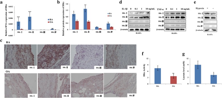
The expression and activity of HKs in synovial tissue from RA patients. a, b HKs mRNA and protein were detected in the synovial tissues from RA (n = 16) and OA (n = 12) patients by real-time PCR and Western blot. The relative expression ratio of HK protein in both groups is shown in the right. c Immunohistochemical staining of RA and OA synovial section using antibodies against HK-I, HK-II, HK-III, and HK-IV. Original magnification × 200. After stimulation with IL-1β (d, left), TNF-α (d, right), and CoCl2 (e, 10 μM)-induced hypoxia for 24 h, total protein was extracted from RASFs (n = 3) and subjected to Western Blot analysis for HK protein. f Hexokinase activity was determined in SFs from RA (n = 6) and OA (n = 6) patients and presented as nanomoles/milligram protein/min. g The lactate levels in the supernatant of RASFs (n = 6) and OASFs (n = 6) were determined by ELISA. Data represent as the mean ± SE. The statistical significance of differences between RA and OA group was determined. *p < 0.05, **p < 0.01, ***p < 0.001significantly different

