
Figure 3.
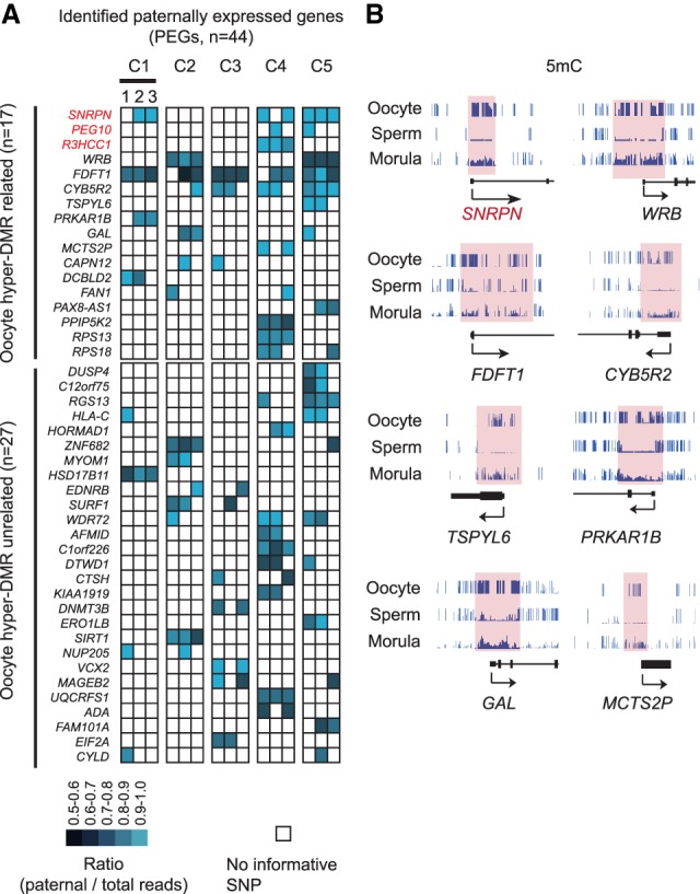
A subset of paternally expressed genes (PEGs) harbor germline differentially methylated regions (DMRs). (A) Heat map showing PEGs identified in this study. Each column represents one morula; a total of three morulae were analyzed for each couple. PEGs with oocyte hyper-DMR within 10 kb of the genes were considered as DMR-related (top panel). The remaining was considered as DMR unrelated (bottom panel). (B) Genome browser view showing examples of PEGs containing oocyte hyper-DMRs at promoter regions. DNA methylation in oocyte, sperm, and morula is shown.