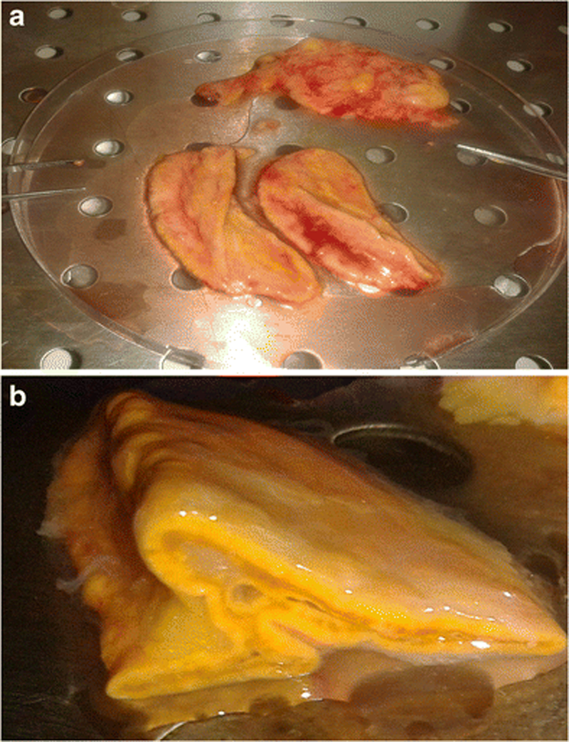
Figure 1. Photographs of human adrenal glands obtained from organ donors.

A) Two whole human adrenal glands in which the surrounding fat has been removed; B) Human adrenal gland sectioned. Note that under the capsule the cortex and medulla are intermingled and there is no separation between them.
